Storyboards for Scientific Animations
I have worked on many storyboards for scientific animations over the past few years. In approaching each project I usually work on gaining a thorough understanding of the concepts presented in the script or the project brief, this includes reading relevant scientific literature, and sourcing microscope imagery, and molecular models. After this initial research stage, I begin to quickly sketch out the complete storyboard in rough thumbnails. The main purpose of this is to allow for rapid ideation and testing out of different visual concepts, but it also contributes greatly to general composition, the motion of characters, and defining the environment in each scene without getting caught in the details of the characters or interactions between them. Once the thumbnails are in a good place I move on to the refined storyboards. Much of my work is in greyscale which allows for a faster production time.
I thoroughly enjoy the process of developing storyboards for scientific animations as it allows me to integrate my innate scientific curiosity as well as continue to develop my artistic skillset. Furthermore, I see the benefit of visual representations in science education as a valuable tool for communicating complicated concepts in ways that a more digestible, clear, and easier to understand.
Examples of a few short storyboard sequences:
1) This set of three panels served to establish a cellular environment that included a tumor mound and immune cells. The camera continues to move through the environment towards the tumor mound, whilst the immune cells interact with tumor cells. There is little immune cell activity due to the immune evasion mechanisms of the tumor cells. By the end of the scene, the camera has approached a few immune cells interacting with some tumor cells, the cellular details visible. The camera blast zooms to a new scene (not shown) at the interface between one of the immune cells and a tumor cell.
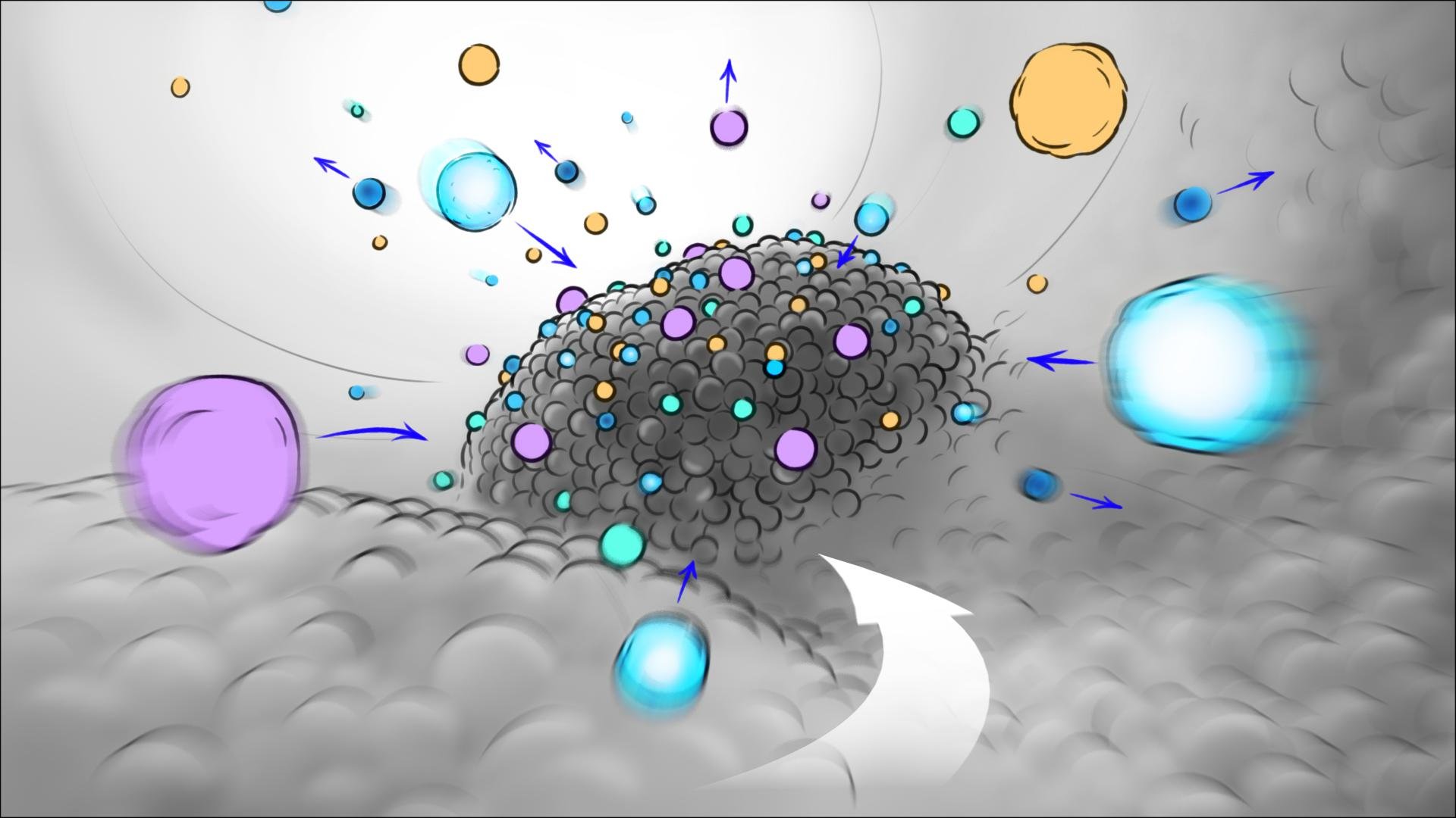
1 Cellular wide

2 Cellular mid

3 Cellular close-up
2) This set of three panels illustrates the transition between three different environments (the surface of a tooth with a thin layer of plaque, the inside of the mouth of someone brushing their teeth, and the initial transition to a non-descript environment in which I introduce the product.

1 Tooth surface

2 Inside a mouth

3 Product environment
3) This set of panels includes the transition between two scenes. The sequence begins with the camera static in a blood vessel with red blood cells pumping through the vessel towards the camera. One of the red blood cells moves right into the camera facilitating the transition to the next scene in which the initial environment motion is matched and quickly decelerates as the camera approaches a hemoglobin protein within the red blood cell. The camera motion in the second scene is utilized to focus the eye on the most important aspects, such as the introduction of hemoglobin, heme, iron, and oxygen.
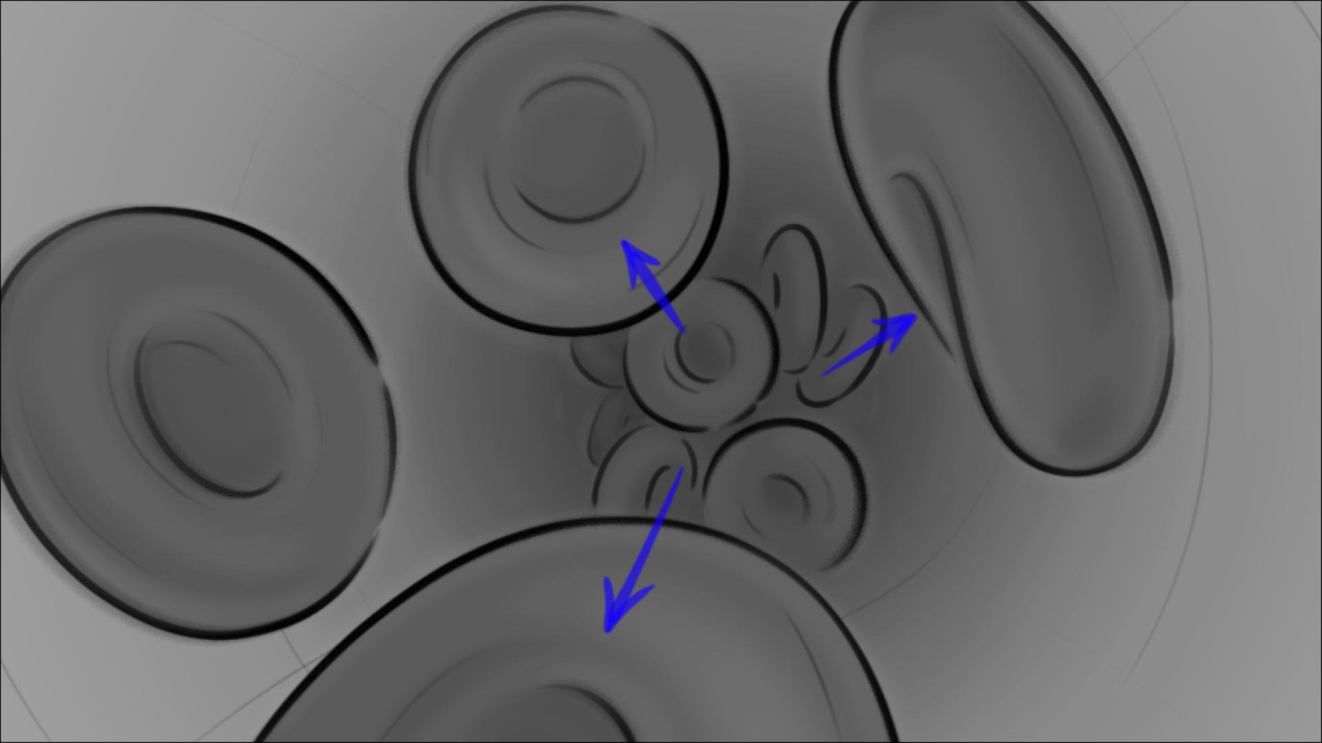
1 Blood flow

2 Blood flow

3 Inside blood cell

4 Inside blood cell

5 Inside blood cell

6 Inside blood cell
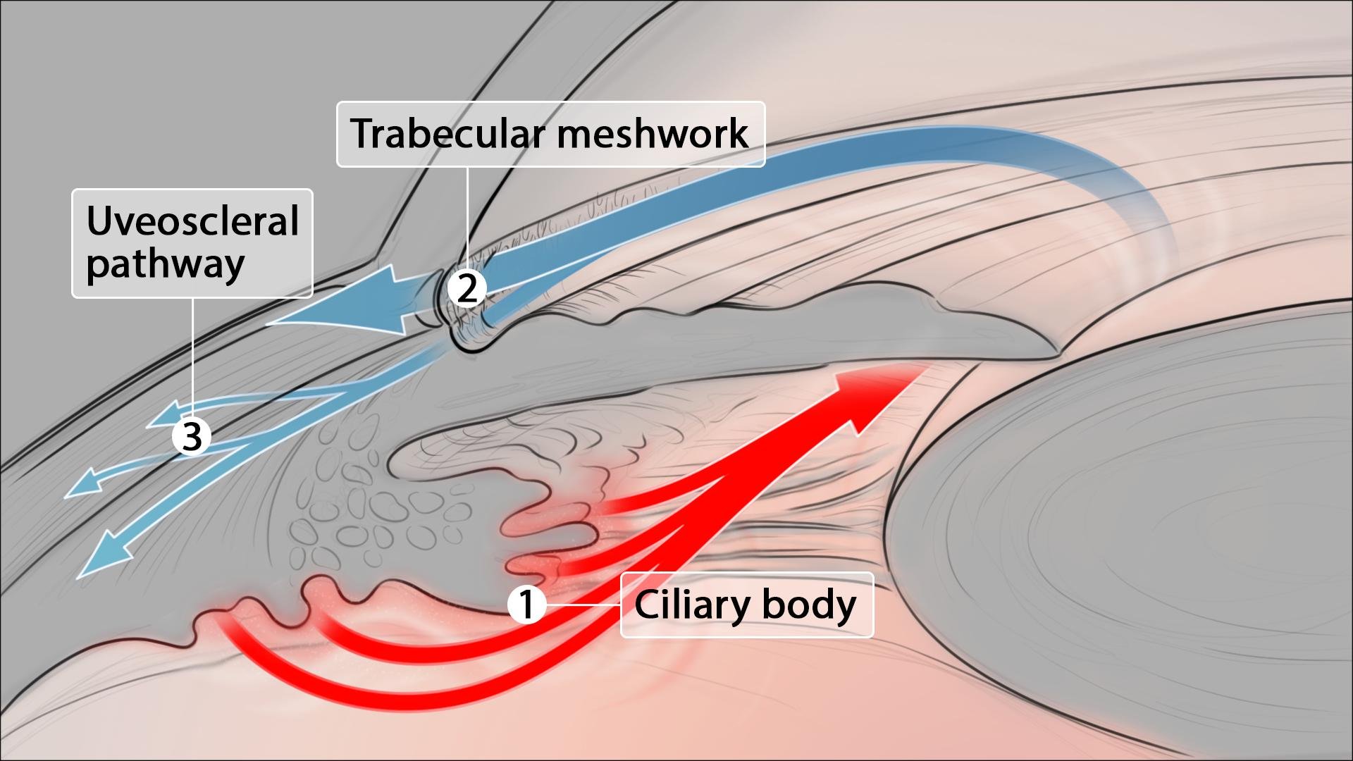
4) This set of panels also includes the transition between two scenes. The first scene is an establishing shot of a human eye, which then transitions to the next scene, a cross-section of the eyeball. The framing of each of the panels was tailored toward communicating the most important visual information without including details that were superfluous to the content being communicated.

1 Eye

2 Eyeball cross-section
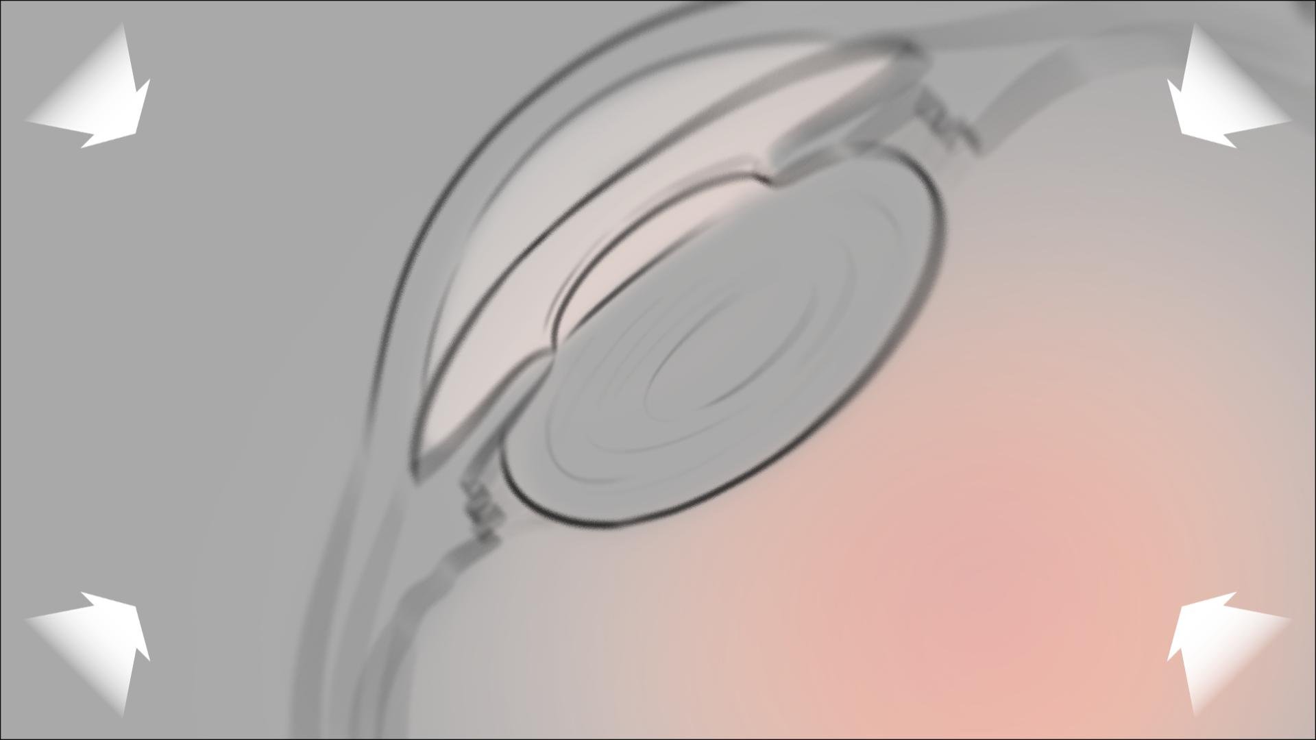
3 Eyeball cross-section mid
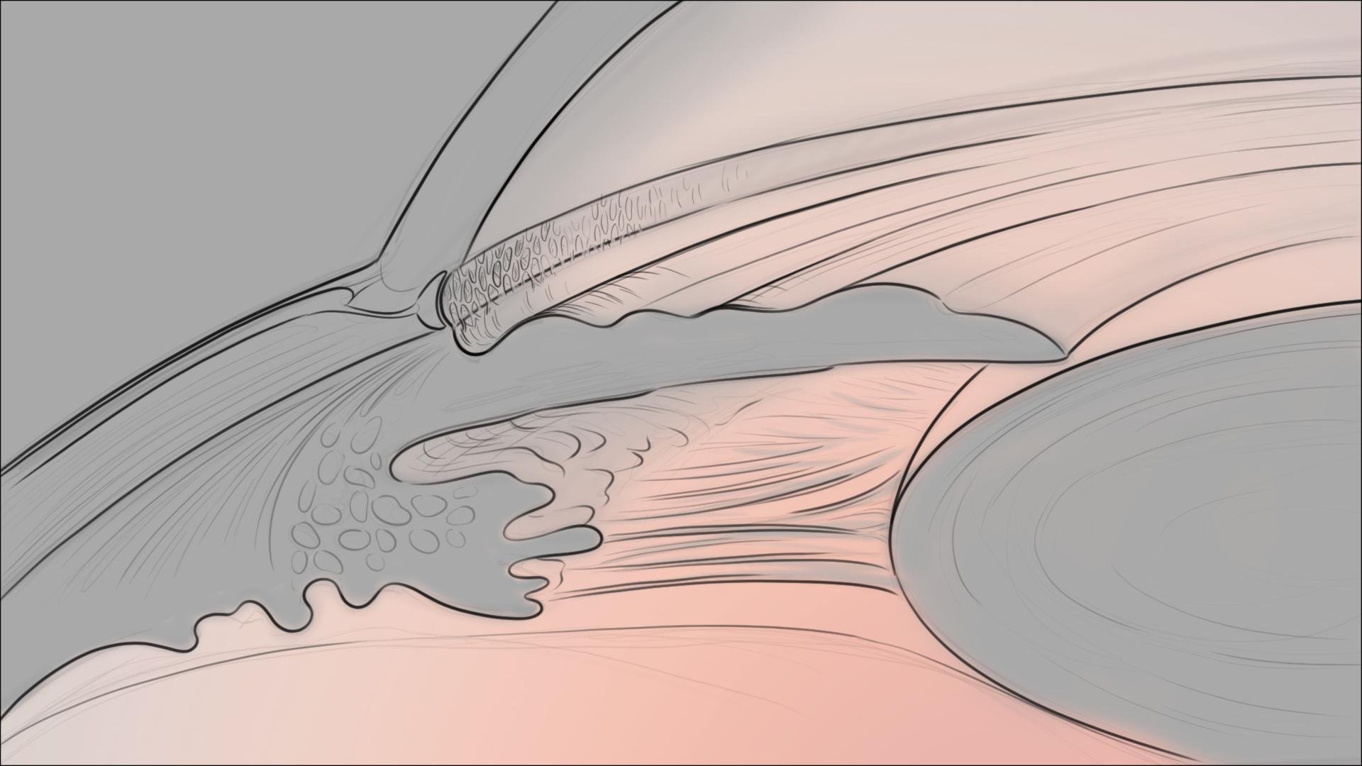
4 Eyeball cross-section close-up

5 Eyeball cross-section close-up
6 Eyeball cross-section close-up
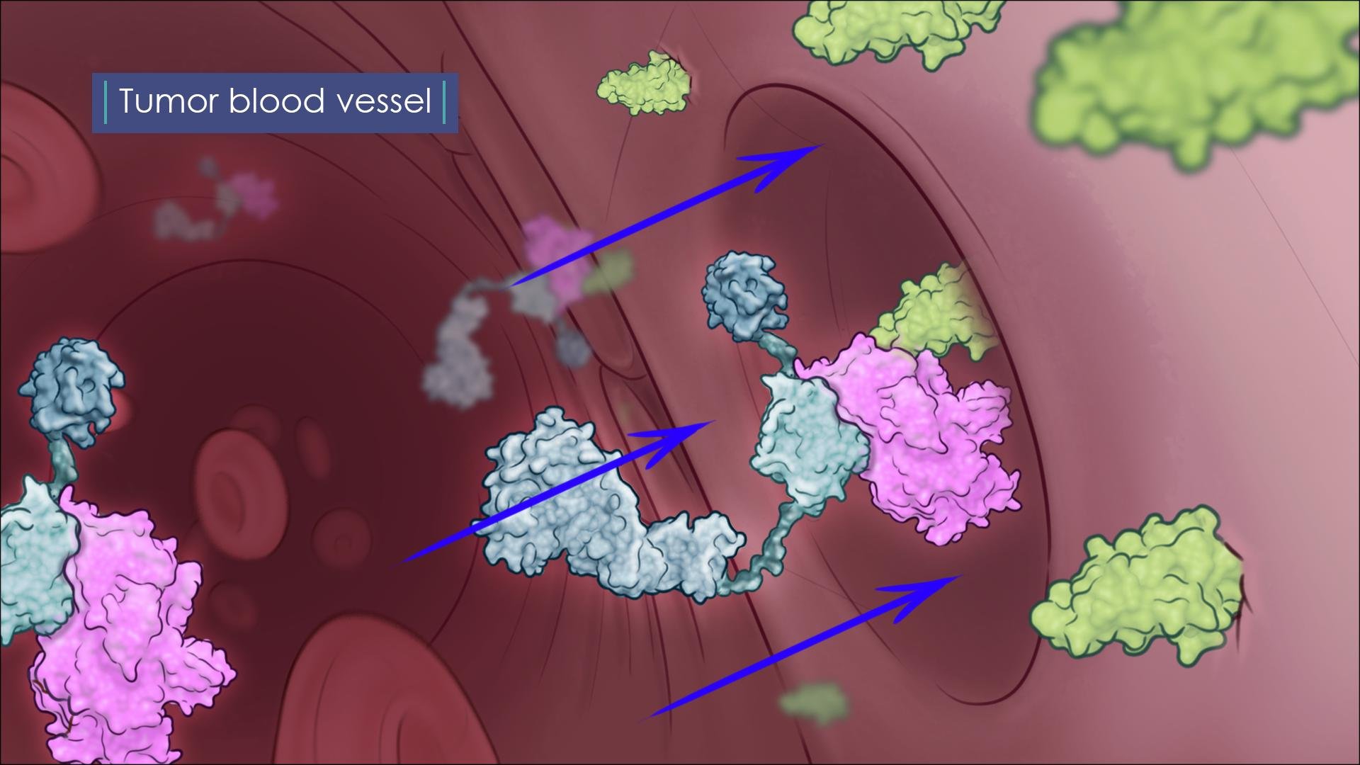
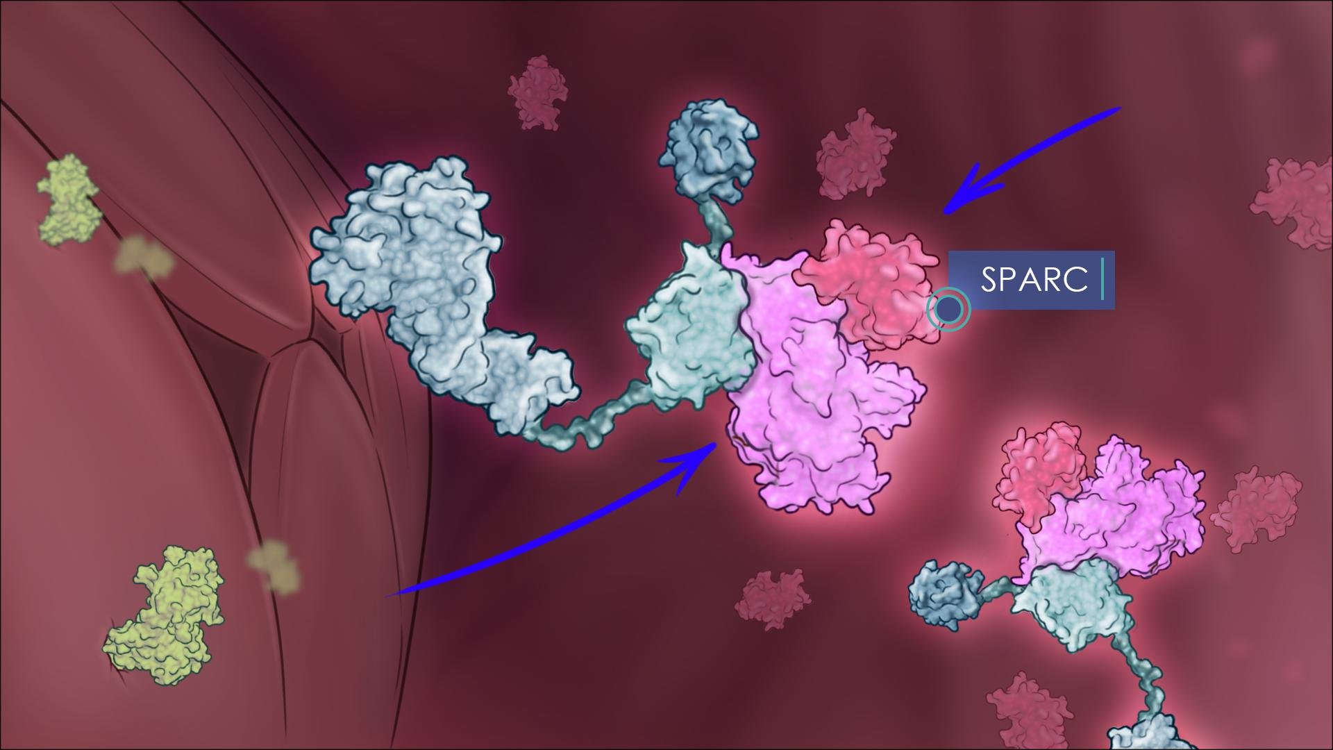
5) This set of panels includes transitions between three scenes. The first scene is within a blood vessel, the main protein shown in teal is interacting with several other proteins and receptors. The sequence transitions from the blood flow environment to the tissue environment surrounding the blood vessel by utilizing an intermediate scene representing transcytosis of the main protein across an endothelial cell.

1 Blood vessel
2 Blood vessel

3 Transcytosis

4 Tissue
5 Tissue
